
Red ring rot is the most important stem decay of conifers in the northern hemisphere. The disease was studied by Robert Hartig in his usual meticulous, insightful manner [5]. Although it is the most studied stem decay, there is still confusion about what the infection courts really are and which lead to most decay. Still, it is considered one of the best examples of a true heart rot.
The disease has also been called “conk rot” [1] because conks are frequently present on infected, living trees. Many stem-decay fungi are not so accommodating.

Hosts
One reason this is such an important disease is the wide host range. Most of the important conifer species of the northern Hemisphere can be infected. Among the most common host genera are Abies, Larix, Picea, Pinus, Pseudotsuga and Tsuga [2]. The featured image above is on Pseudotsuga menziesii.
Pathogen


Porodaedalea (Phellinus) pini is the traditionally recognized pathogen. In English, some call it the ring-scale fungus [1]; in German it is Kiefernbaumschwamm [5] or Kiefernfeuerschwamm [3]. It is really a complex of species. It is surprising that the taxonomy of the most important stem decay pathogen in the world is still far from settled. I won’t drag you through all the gory details; if you’re interested see this blog post. Here we’ll just consider the current situation as we know it:
- Porodaedalea pini and P. chrysoloma, a second species that usually has been recognized in North America (NA), occur in Eurasia but apparently not in NA.
- P. cancriformans, causing cankers on Abies spp. in western NA, appears to be a good species but could use more work.
- There may be two undescribed phylogenetic species of Porodaedalea in southeastern and southwestern NA.
- Porodaedalea piceina is the most common and widespread species in NA and also occurs in northern Europe (often called P. laricis there).
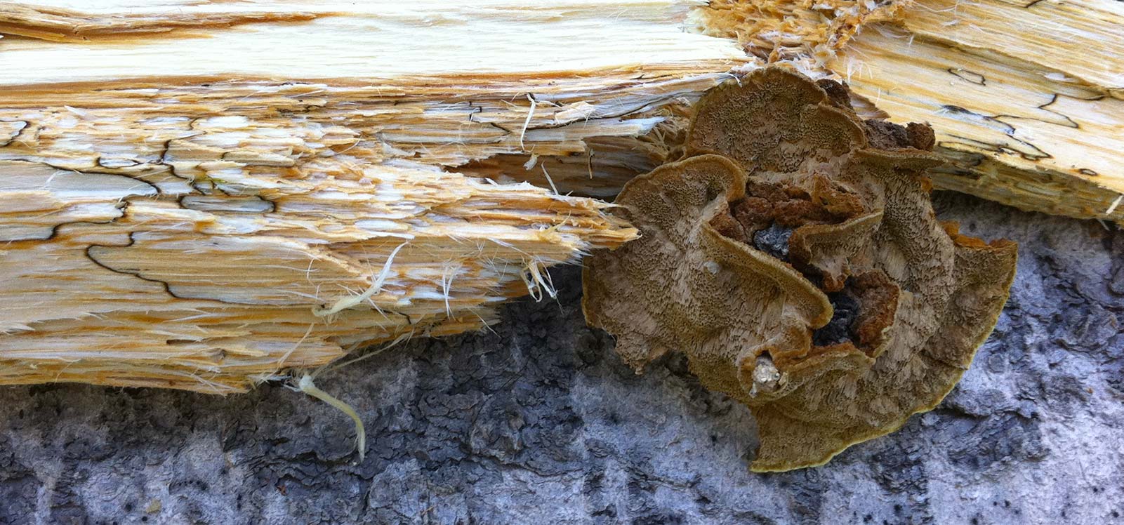
In most species, conks may be large (~10×15 cm and 10 cm tall), solitary or grouped, ungulate to triquetrous (hoof-shaped to triangular in profile), but they may be thin, shell-shaped, densely grouped and imbricate (with overlapping, fused pilei). The pileus surface is light red-brown to almost black, zonate, and hairy near the margin to hairless and encrusted. The pore surface is yellow-brown. Pore shape is variable even within species, ranging from circular to angular to daedaleoid (labyrinthine, maze-like). Context is red to yellow-brown. Tube layers are thin (up to 6 mm thick) and indistinct from one another.
Environment

In an enormous study of red ring rot in Pseudotsuga menziesii (Douglas-fir), Boyce & Wagg found that, on good sites, decay begins at a younger age and reaches higher levels than on poor sites [1]. They also found that, on a given site, fast-growing trees are more susceptible to infection than slow-growing trees.
The disease is more common in central and southern Oregon (dry) than in other parts of the Pacific Northwest; also more common on southern slopes than northern [1]. The higher temperatures may be more important than moisture in these cases.
Disease Cycle
Infection originating at large branch stubs in the mid-trunk section of older trees has long been recognized, and that it is of common occurrence cannot be doubted.
W.R. Haddow (1938)
Infection court. As with the identity of the pathogen species, it is amazing that knowledge of the infection court is confused for the most important stem decay in the world. The introductory page on stem decays has an extensive discussion of infection courts, much of it dealing with red ring rot. Suffice it here to say that study after study has concluded that wounds and branch stubs are important infection courts (see [8] and references therein). On the other hand, there is one study indicating alternative infection courts, small dead twigs and weevil-killed leaders [4], and even its author acknowledged that the other infection courts were undoubtedly common (see quote). Yet, in recent years, some have suggested that Porodaedalea spp. do not infect appreciably through wounds and branch stubs, sweeping aside all the evidence to the contrary [7]. Clearly more work on the relative importance of various infection courts is needed.
Distribution in the tree
Decay columns are more or less centered at the infection courts, because growth up and down is roughly the same. When trees are young, the infection courts are not far from the ground. As trees grow larger and older, the infection courts are higher, and resulting decay columns are as well. Thus, in old stands, where the trees infected the longest are already dead, decay columns tend to be relatively high in the trees [1].
However it gets in, the pathogen initially establishes in the heartwood. It then grows much faster longitudinally than radially, and decay columns get longer over time. At the low end, I have found it extending into the root collar and major roots of Picea engelmannii on several occasions, even bumping into Armillaria solidipes there. See the gallery below for a peek at the party they had!


Columns of decay do not have neat, conical ends as do those of some other stem decays [1]. At the column ends, cross-sections reveal the typical ring-like arcs of stain (incipient decay) and advanced decay, while vertical sections show streaks and spires with areas of sound wood between. Away from the ends of the column, incipient decay extends radially 5-7 cm out from advanced decay [1]. Vertically, it may extend an average of 1 m from advanced decay and rarely as far as 9 m.
Sapwood is often attacked after development of decay in the heartwood, as with many stem-decay fungi (see two sections of the main Stem Decays page: True stem decays vs. wound decays and Invasion of sapwood). In Pseudotsuga menziesii, sapwood is usually narrowed uniformly, but in some cases triangular outgrowths of the decay approach or reach the cambium [1]. In Pinus strobus, red ring rot often results in thin sheaths of living sapwood as the inner sapwood is killed, invaded, and decayed [4]. In Abies spp., P. cancriformans kills large areas of sapwood and cambium, forming large cankers on which it fruits [6].
Fruiting
The requirements for and functions of fruiting generally follow the typical stem-decay disease cycle. In some areas and hosts, fruiting occurs more consistently than for many stem decays, and is thus a more useful indicator of infection [1].
Symptoms and Signs
Punk knot. This is one of the best symptoms, because it occurs frequently and is diagnostic. This begins as growth of the fungus from the inside through the sapwood along the trace of a dead branch. When it reaches the surface, the branch trace is first surrounded by red-brown (often called golden brown) fungal mycelium, which looks just like the mycelium that makes up the context above the tube layers of a conk. The fungus attacks the cambium and phloem of the stem around the stub. This may stimulate the cambium to produce more cells than usual, causing swelling around the punk knot. Alternatively, the cambium may be killed around the knot, which results in a sunken punk knot as the stem grows where it remains alive.
Another symptomatic host reaction associated with punk knots is resinosis. This may be moderate and difficult to distinguish from normal resin exudation in some species, such as Picea engelmannii. In other species, such as Pinus strobus, resinosis is a useful symptom when it occurs copiously at punk knots and to a more limited extent in the absence of disease. It can even occur around knots and stubs that are not punky, but are near stem infections [4].
The fungus also slowly decays the branch stub. When the knot decays more or less completely, it is usually replaced by fungal mycelium, but it can leave a hollow core (‘punk cavity’). In either case, as the tree grows in diameter and buries the end of the stub, the fungus continues to grow radially as a cylinder of mycelium beyond the stub, and continues to subdue the surrounding cambium to prevent overgrowth by callus tissue.
Punk knots can be neither sunken nor swollen, and without much resinosis. But shaving the bark with a hatchet would reveal the mycelium (which is rarely visible otherwise). Another good way to detect the disease is tugging and twisting small dead branches within reaching distance. If the disease is there, often those branches will easily pull out along with a piece of the branch trace with the characteristic mycelium color.


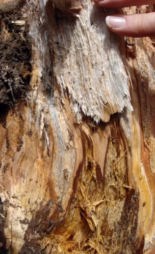
Decay. In some cases the decayed wood may be visible in snapped trees, felled trees, dead branches, etc. The characteristic decay features can go a long way to narrowing the identity of the pathogen. The decay is a white pocket rot, sometimes referred to as white speck. It begins as a red stain in longitudinal streaks, often as a short arc following the annual rings (thus the disease name). As decay becomes noticeable, white pockets develop in the wood. They are elliptical to fusiform, with the long axis oriented longitudinally.
Signs of the disease include conks (basidiomata) and the mycelium in punk knots. On infected trees, fruiting may range from rare to somewhat common, so absence of conks is not informative. In P. strobus in northern Ontario, only 1 out of 50 infected trees may have conks, and small ones at that [4]. Conks are not formed directly on live bark. On live trees, they are most common subtending old branch stubs, but also occur on wounds and cankers. In some cases they are produced more abundantly on dead trees than on live. Haddow found them much more abundantly on fallen trees and cull logs than on live Pinus strobus [4] and the same is often true of Picea engelmannii in the Rocky Mountains. Conks vary from a few cm to 50 cm wide, but size may be less variable in a given host and region.
Distribution
Although the pathogen species vary, the disease occurs throughout the northern hemisphere.
Damage and Management
See the general Stem Decays page for information on impacts and management.
- 1.Boyce JS, Wagg JWB. 1953. Conk Rot of Old-Growth Douglas-fir in Western Oregon. Bulletin 4. Oregon Forest Products Laboratory and Oregon State Forestry Department <https://ir.library.oregonstate.edu/downloads/k35695737>.
- 2.Brazee NJ, Lindner DL. 2013. Unravelling the Phellinus pini s.l. complex in North America: a multilocus phylogeny and differentiation analysis of Porodaedalea. Forest Pathology 43:132–143 <https://research.fs.usda.gov/treesearch/56263>.
- 3.Butin H. 1983. Krankheiten der Wald- und Parkbäume: Leitfaden zum Bestimmen von Baumkrankheiten. Stuttgart: Georg Thieme Verlag.
- 4.Haddow W. 1938. The disease caused by Trametes pini (Thore) Fries in white pine (Pinus strobus L.). Transactions of the Royal Canadian Institute 22(1):21–80.
- 5.Hartig R. 1874. Wichtige Krankheiten der Waldbäume. Beiträge zur Mycologie und Phytopathologie für Botaniker und Forstmänner. Berlin, Heidelberg: J. Springer Berlin Heidelberg <https://link.springer.com/book/10.1007%2F978-3-642-50830-1>.
- 6.Larsen MJ, Lombard FF, Aho PE. 1979. A new variety of Phellinus pini associated with cankers and decay in white firs in southwestern Oregon and northern California. Canadian Journal of Forest Research 9(1):31–38 <10.1139/x79-006>.
- 7.Vasaitis R, Gonthier P, Nicolotti G. 2013. Heart rots, sap rots and canker rots. In: Infectious Forest Diseases, pp. 197–229. Wallingford: CABI <https://www.scribd.com/document/544566959/10-Heart-Rots-Sap-Rots-and-Canker-Rots-PDFDrive>.
- 8.Wagener WW, Davidson RW. 1954. Heart rots in living trees. The Botanical Review 20(2):61–134 <https://www.jstor.org/stable/4353511>.